Psoriasis is a disease that most people think of as primarily a skin disease because the condition causes a persistent rash in various areas of the body. Psoriatic arthritis is a type of joint disease that occurs in roughly seven percent of people who have psoriasis. Psoriatic arthritis affects people of all ages, but most get it between the ages of 30 and 50. Usually a patient has psoriasis (the skin rash) for many years before the arthritis develops, and the arthritis comes on slowly. But this is not always the case. No matter what, patients with psoriatic arthritis must manage both the outbreaks of itchy, scaly skin and the pain and stiffness of arthritis.
http://www.eorthopod.com/content/psoriatic-arthritis
Sunday, March 27, 2011

Saturday, March 26, 2011
OTHER ARTICULAR MANIFESTATIONS IN PSORIATIC ARTHRITIS
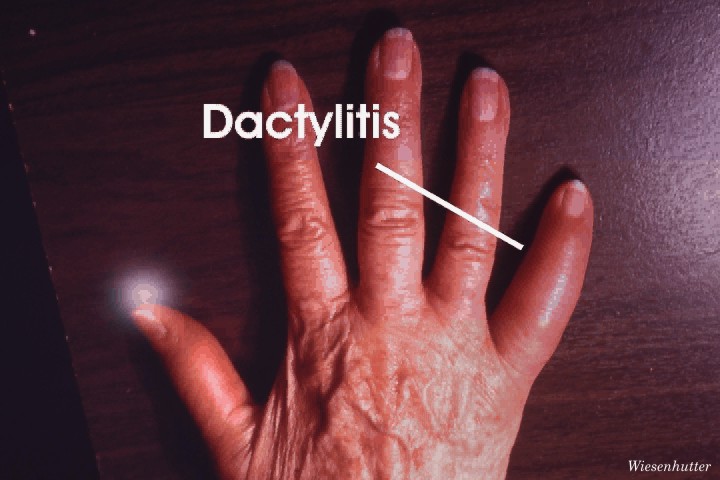
Dactylitis
Dactylitis, or sausage digit, is a typical feature of PsA. It refers to infl ammation of the whole digit. It likely
results from both synovitis in the joints of the digit, as well as tenosynovitis, particularly in the fl exor tendons.
Dactylitis most commonly affects the toes, but fingers are affected as well.
It should be noted that dactylitis may become chronic, such that it is no longer painful or red, but remains as a chronically swollen digit, which may not respond to therapeutic intervention.
Dactylitis http://www.cdaarthritis.com/images_slides/43_spon2_d_adactylitisfinger_720.jpg
Tenosynovitis
Tendonitis or tenosynovitis occurs frequently among patients with PsA. Infl ammation may affect the fl exortendons of the fingers, as well as the extensor carpi ulnaris, sites that are commonly affected in RA. Achillestendonitis is commonly seen, as is plantar fascitis. These may interfere with function and may lead to disability. In PsA, tendonitis may be associated with tendon nodules and signifi cant functional limitation.

Enthesitis
Infl ammation of the enthesis, site of insertion of tendoninto bone, is another typical feature of PsA. Enthesitis
may occur at any tendon insertion site, but most commonlyaffects the plantar fascia, Achilles tendon insertion,
insertion of tendons at the knee and shoulder, aswell as the pelvic bones. It has been suggested thatenthesitis alone in the presence of psoriasis may be sufficient for the diagnosis of PsA.

Other Extra-Articular
Manifestations
Iritis is an extra-articular feature common to all spondyloarthropathies and is also seen among patients with
PsA. Some 7% of patients with PsA present with iritis,and it can also be seen among patients with psoriasis
without arthritis.
Urethritis is also a feature of seronegative disease. It
is less common in PsA than in the other members of the
spondyloarthritis group.
Bowel involvement may occur in patients with PsA
and is usually nonspecifi c colitis.
Cardiac abnormalities have been reported among
patients with PsA, including dilatation of the base of
the aortic arch which occurs in ankylosing spondylitis.
More recently it has been recognized that patients
with PsA are at risk for cardiovascular disease.
This may be related to the metabolic abnormalities
associated with PsA, including hyperlipidemia, hyperuricemia,
as well as lifestyle factors such as obesity and
smoking.
X-Ray Findings in Psoriatic Arthritis
The general features and diagnostic signs of psoriatic arthritis include: asymmetric distribution, soft tissue swelling, normal bone mineralization, erosions, fluffy periostitis, and a narrowed or widened joint space.
In the hands, psoriatic arthritis is usually asymmetric. A ray pattern is usually seen in the DIP joints. Mouse ears sign, pencil in cup deformity, and opera glass hand deformity are also radiographic signs of psoriatic arthritis in the hands and feet. Joint space widening is common due to fibrous tissue deposition and bone destruction.
The sacroiliac joints are involved in up to 50% of patients with psoriatic arthritis. The distribution is usually bilateral, but asymmetric and the joint space is narrowed.
Psoriatic arthritis in the spine is most common in the upper lumbar and lower thoracic area. There is a presence of marginal and non-marginal syndesmophytes, with the latter being the most common. It is usually asymmetric and unilateral.
*All information gathered from Yochum and Rowe's Essentials of Skeletal Radiology - 3rd Edition.
In the hands, psoriatic arthritis is usually asymmetric. A ray pattern is usually seen in the DIP joints. Mouse ears sign, pencil in cup deformity, and opera glass hand deformity are also radiographic signs of psoriatic arthritis in the hands and feet. Joint space widening is common due to fibrous tissue deposition and bone destruction.
The sacroiliac joints are involved in up to 50% of patients with psoriatic arthritis. The distribution is usually bilateral, but asymmetric and the joint space is narrowed.
Psoriatic arthritis in the spine is most common in the upper lumbar and lower thoracic area. There is a presence of marginal and non-marginal syndesmophytes, with the latter being the most common. It is usually asymmetric and unilateral.
*All information gathered from Yochum and Rowe's Essentials of Skeletal Radiology - 3rd Edition.

Anteroposterior radiograph of the hands shows subchondral erosions of the fourth left distal interphalangeal joint and right third and fourth proximal interphalangeal joints with periosteal reaction. Courtesy of Bruce M. Rothschild, MD

Anteroposterior radiograph of the feet shows arthritis mutilans. Courtesy of Bruce M. Rothschild, MD.

Anteroposterior radiograph of the thumb shows arthritis mutilans. Courtesy of Bruce M. Rothschild, MD.

Anteroposterior radiograph of the pelvis in a male patient shows sacroiliitis in association with psoriasis. Courtesy of Ali Nawaz Khan, MBBS.

Left, lateral radiograph of the dorsal spine of a 38-year-old man shows spondylitis in association with psoriasis. Note the squaring of the anterior vertebral bodies. Right, radiograph of the pelvis in the same patient shows sacroiliitis and erosive changes around both hip joints. Courtesy of Ali Nawaz Khan, MBBS.
X-Ray Finding in Psoriatic Arthritis
There are five different types of psoriatic arthritis which include:

- Arthritis mainly in the small digits of the fingers or toes (55-70%)
- Asymmetrical arthritis in the joints of the extremities (30-50%)
- Symmectrical polyarthritis which can look like rheumatoid arthritis (15-70%)
- Arthritis mutilans: this is very rare, but can be very destructive to the joints (3-5%)
- Arthritis in the sacroiliac joints and the spine (5-33%)
Posteroanterior radiograph of the hands shows wrist fusion. Courtesy of Bruce M. Rothschild, MD.
X-Ray Finding in Psoriatic Arthritis
Twenty-three percent of people with arthritis have psoriatic arthritis. Psoriatic arthritis usually occurs in people 30-50 years old, and usually about 10 years after the first signs of psoriasis occur. It is very rare to develop psoriatic arthritis without first having psoriaris.

Anteroposterior radiograph of the abdomen shows fusion of the sacroiliac joints. Courtesy of Bruce M. Rothschild, MD.
Therapeutic approach to Psoriasis
The following has been shown to help psoriatic patients: decreasing sugar, meat, animal fats, and alcohol; increase dietary fiber, and cold water fish, have a healthy B.M.I.; eliminate gluten and any other potential food alergies.
Supplements shown to help the psoriatic patient include: multi-vitamin, mineral formula, flaxseed oil, vitamin A, Vitamin D, Vitamin E, Chromium, selenium, zinc.
Botanical Medicines that may be useful include: Goldenseal, smilax sarsparilla , and milk thistle
Reducing stress levels have been shown to help psoraitic patients.
Justin Felsman
Supplements shown to help the psoriatic patient include: multi-vitamin, mineral formula, flaxseed oil, vitamin A, Vitamin D, Vitamin E, Chromium, selenium, zinc.
Botanical Medicines that may be useful include: Goldenseal, smilax sarsparilla , and milk thistle
Reducing stress levels have been shown to help psoraitic patients.
Justin Felsman
Subscribe to:
Posts (Atom)